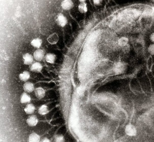
How do scientists detect viruses? Being sub-microscopic entities, it would be better to say them nanoscopic (their size varies between 30 nm and 500 nm, where 1 nm is a million times smaller than 1 mm – the new coronavirus, SARS-CoV-2 , is about 100 nm), it is not strange to think that its detection by “simple” observation under an optical microscope is very difficult.
Only with high-resolution optical microscopes, with a high magnification capacity, it is possible to observe viral particles. In fact, viral architecture was only better known with the advent of the electron microscope, and it was with it that virologists, that is, those specifically dedicated to the study of viruses, captured the profile of these pathogens.
It was precisely by microscopic observation in 1965 of the profile of viruses that resembled a crown that the term coronavirus was assigned to this viral family.
On the other hand, X-ray diffraction crystallography (a technique used to resolve molecular structures) allowed us to detail the spatial regularity present in the protein capsules (capsids) that form viruses.
The first virus to be crystallized, in 1935, and to be studied in detail by X-ray diffraction, in 1941, was the tobacco mosaic virus.
Interestingly, it was Rosalind Franklin (1920 – 1958) – the least known and somewhat ignored researcher of the structure of DNA – who solved the three-dimensional structure for this virus in 1955.
The “visualization” by these techniques has the “inconvenience” of not allowing the virus to be kept in a living cellular environment: when preparing the sample to be observed, the researcher has to “definitely” immobilize or crystallize a moment of the virus's life. What is gained in form detail is lost in the dynamics of viral activity.
Back to the optician: with this microscope it is possible to keep biological preparations alive and thus visualize, with the necessary patience and expertise, the strategies, movements and viral damage. Let's say the two views complement each other.
This difficulty in visually detecting viruses explains why the first viral images were only obtained without distinction in the middle of the last century.
In 1938, B. Borries, H. Ruska and E. Ruska (the latter was awarded the Nobel Prize in Physics in 1986 for the development of the electron microscope) presented to the world the first electron micrograph of viruses, more precisely of viruses ectraomelia e vaccinia (responsible for types of smallpox), confirming the physical existence of a pathogenic entity long announced and fought.
It should be remembered, as a curiosity, that the first microorganisms were visualized with an optical microscope, in the year 1668, by Antoine van Leeuwenhoek (1632 – 1723).
From what has been said, it is suspected that the first investigations into the existence of the non-cellular pathogenic entity responsible for fatal diseases, with enormous capacity to spread by contagion and infection, must have been carried out by means of investigation other than microscopy.
In fact, the laboratory work of identifying and isolating the virulent entity present in filtered biological fluids, animal sera or bacterial suspensions was a Herculean task whose story deserves to be told.
The past of viruses
When someone suffers a physical aggression or a tragic fall, it is unquestionable to associate the disease state, more or less severe or even fatal, that comes with it, with the wound caused by the enemy's sword or the fracture that may be exposed in the leg after a tumble.
But when someone is prostrate overnight, burning with fever and with the body covered with rashes and redness, without anyone or anything detectable by eye, auditory or olfactory witnesses being identified as the direct cause of the disease, then the human imagination burns a divine wrath, an enemy plague, an envious evil eye, among other metaphysical curses.
In fact, if it is against common sense to understand that the Earth revolves around the Sun and not the other way around, just with our unarmed gaze and without using any lenses, it is not surprising that it is against sense to attribute to organisms invisible to our eyes the cause of countless diseases.
And so, for a long time, the blame for the origin and spread of the plague fell on the infected host (the sick person) and not on the microorganism that infected it, because it found the ideal conditions to develop in it.
We know that Hippocrates of Cos (460-370 BC), the Greek considered the father of medicine, around 400 BC made epidemiological observations of many diseases, despite not having attributed to them any organic causative agent.
Many centuries later, in 1546, Girolamo Fracastoro (1478-1553) proposed the theory that epidemic diseases (that is, diseases that develop in one place quickly and claim many victims in a short period of time) are contagious and spread through tiny particles and over long distances.
But, the first association between a disease and an infectious organism specific to it that we know to have been carried out experimentally, was made in 1863 by the French physician Casimir Devaine (1812 – 1882). The disease in question was anthrax, or anthrax, and the causative microorganism a bacterium, the Bacillus anthracis.
However, the demonstration, methodologically scientific through controlled experiments, that this bacterium is in fact the agent, or the pathogen, causing that pathology, was only carried out in 1876 by Robert Koch (1843 – 1910) and Louis Pasteur (1822-1895 ) – the latter the founder of microbiology.
We owe to these two scientists, among others, the proposal, demonstration and dissemination of the theory that proposes that microorganisms are the cause of countless diseases, putting an end to the theory of spontaneous generation of disease (and life!), first shaken in 1668 , for the beautiful scientifically controlled experiment of Francesco Redi (1626 – 1697).
But let's go back to viruses and answer the question: when did the first evidence appear that there were diseases that, not being caused by bacteria and not generating spontaneously, would be caused by a hitherto undetected agent?
Let us remember that advances in optical microscopy, at the end of the XNUMXth century, allowed the visual detection of bacteria but not viruses.
Koch and Pasteur were able to demonstrate the presence of bacteria in the liquids with which they inoculated the animals, which, as a result, became ill. By filtering these liquids containing bacteria so that the filtrate did not contain them (which they could also confirm and demonstrate with the microscope) and if they inoculated animals of the same species with this preparation, then, if they did not get sick, they demonstrated that the bacteria were the pathogens.
But, in 1892, an observation intrigued the scientific community (and not only!). Russian scientist Dimitri Ivanovski (1864-1920) demonstrated that a disease that afflicted the tobacco plant, tobacco mosaic disease, could be caused by the “simple” contact of the leaves of a healthy plant with the liquid resulting from filtering the extract. of diseased leaves crushed, through a Chamberland filter (porous porcelain filter also called Pasteur) which had pores small enough to prevent the passage of microorganisms then known.
In other words, Ivanovski showed the world that a “filterable agent”, smaller than bacteria, was responsible for triggering a disease in plants.
In 1898, the German Martinus Beijerinck (1851-1931) repeated the previous experience and independently confirmed the existence of something causing the disease in solutions without any bacteria.
Designated this agent by the Latin expression contagium vivum fluidum (living fluid germ) and reintroduced in this context the word virus (also of Latin origin and meaning toxin, poison).
The debate about the nature of the filterable agent then fueled heated discussions: would it be a “living fluid”, an infectious “particle”, or a toxin?
In that same year of 1898, a second similar finding was made in animals. The Germans Friedrich Loeffler (1852-1915) and Paul Frosch (1860-1928), who worked with Koch, filtered a liquid containing the agent of foot-and-mouth disease (which we now know to be a virus of the genus aphthovirus) through a Chamberland filter and showed that the filtrate continued to cause disease.
However, when passing the same filtrate through a fine-grained Kitasato filter (which has much finer pores) they found that the potential to induce infection had remained in the filter.
With this experiment, they had not only demonstrated that the infectious agent was not liquid in nature, but composed of particles, but they had identified for the first time a way to isolate a virus that infects vertebrates.
Furthermore, they showed that the retained agent was somehow capable of replicating. These discoveries mark the beginning of virology as a scientific discipline.
These episodes of discovering the nature of viruses illustrate well the meaning of what we understand by scientific discovery. Although Ivanovsky was the first to observe the existence of something passing through a filter and causing disease, all his publications show that he did not understand that his observations implied the existence of a pathogenic microbe distinct from bacteria.
Beijerinck, for his part, was convinced of the existence of something other than bacteria, but he always maintained that it had a fluid and non-corpuscular nature.
Only Loeffler and Frosch enunciated a set of hypotheses and planned controlled experiments in order to be able to conclude, without misunderstandings, on the existence of a new agent, with sub-microscopic dimensions, capable of inducing disease and replicating itself: viruses.
In fact, good science is not just a collection of good observations and new data. It always needs new evidence to be processed by creative and critical thinking that allows scientists to make an impartial and correct interpretation of their experimental findings and reach conclusions verifiable by all.
Author António Piedade is a Biochemist and Science Communicator.
Science in the Regional Press – Ciência Viva


Comments