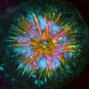
 A study by a team from the Institute of Molecular and Cell Biology (IBMC), University of Porto, published in the Journal of Cell Biology, presents a set of unique images of the end of immortal cancer cells.
The death of these cells results from the alteration of a small two-protein binding component (PLK1 to CLASP2), which disrupts cell division. With this work, the IBMC team of researchers presents a new molecular regulation mechanism and opens up a whole field for new anti-tumor therapeutic approaches, already under patent application.
A team from the Institute of Molecular and Cell Biology (IBMC, University of Porto) genetically manipulated the area of Plk1 binding to CLASP 2 in cell lines. The researchers used an immortalized tumor line (Hela cells) that has served as a model for many studies since 1951, when it was isolated.
“By preventing the binding of these proteins, we prevented a simple chemical reaction of CLASP2 phosphorylation from taking place and the result is the death of the affected cells”, explains Hélder Maiato, coordinator of the team. “It is interesting to realize that controlling a simple phosphorylation reaction can lead to such a dramatic event for a cell”, adds the researcher.
Although the explanatory model is simple, preventing a chemical reaction in such a specific location in the cell involves great technical complexity. The researchers had to prove that by disabling the phosphorylation of CLASP by Plk1 the bipolar mitotic spindle, essential to cell division, could not be maintained.
 As Rita Maia, first author of the article, explains, "during division, the cells form a spindle to ensure the equal distribution of genetic material between the two daughter cells", which is a "very coordinated process in order to avoid production of genetic errors, in which CLASSP2 plays a fundamental role”.
When the binding of Plk1 to CLASP2 is prevented, the coordination of the process is affected, the bipolar structure of the spindle is compromised, and the cells simply die. "Instead of two, none!"
And it is precisely the moment of anomalous arrangement of the mitotic spindle that is offered to the researchers' cameras in images comparable to events in astronomy and the cosmos, as can be seen in the figure on the side. It is usual to be presented with images of these in astrophysics studies, but the microcosm of cells manage to offer moments of very equivalent beauty.
“Perhaps the images are spectacular for many, but for us, the most important thing is the perspectives that this study opens up”, says Hélder Maiato.
 For the team involved, the great advance achieved in this study is "the exciting possibility of administering genetically modified Plk1 substrates, which lead to the death of tumor cells and can be used as an alternative therapeutic for certain cancers", as read in the article.
This study has already allowed the elaboration of a patent application regarding the administration of modified CLASP2 for therapeutic purposes.
Author Julio Borlido Santos (IBMC)
Science in the Regional Press – Ciência Viva
Figure: Hela cells in full division. In blue the chromosomes, in green the altered protein, in red the mitotic spindle. This cell has a monopolar structure that will make division impossible and will eventually die.
Original article reference:
Ana RR Maia, Zaira Garcia, Lilian Kabeche, Marin Barisic, Stefano Maffini, Sandra Macedo-Ribeiro, Iain M. Cheeseman, Duane A. Compton, Irina Kaverina and Helder Maiato. Cdk1 and Plk1 mediated a CLASP2 phospho-switch that stabilizes kinetochore–microtubule attachments. J. Cell Biology (2012) 199, 2, 285-301. doi:10.1083/jcb.201203091
http://jcb.rupress.org/content/199/2/285.abstract


Comments